By Dr. Jaday Garcia, PhD, BCDNM, 2026 Global Recognition Award Winner
Last Updated: July 7, 2026
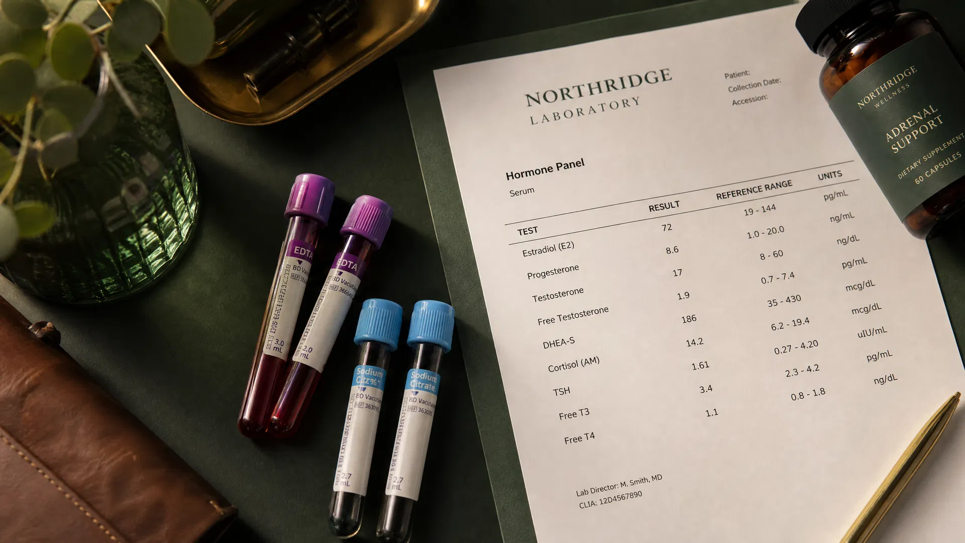
You went to your doctor. You told her you were exhausted, that your brain felt foggy, that your hair was thinning, that your sleep was broken, that your weight had shifted even though nothing in your routine had changed. She listened. She ordered labs. And a week later, you got the call: "Everything looks normal."
But you are not normal. You know it. Your body knows it. And the disconnect between what you feel and what the lab report says is one of the most frustrating experiences in modern medicine.
Here is what I want you to understand: the problem is not that your body is lying to you. The problem is that the labs your doctor ordered were never designed to find what is actually wrong.
I have spent years working with women who arrive at my practice after being told their labs are normal. They are not imagining their symptoms. They are not stressed. They are not aging prematurely. They are experiencing real physiological dysfunction that standard lab testing was never built to detect.
This article will walk you through three specific mechanisms that explain why standard labs miss what is actually happening in your body. I will show you the markers that matter, the ranges that actually reflect optimal function, and the testing methods that give us the full picture. By the end, you will understand exactly why "normal" labs do not mean you are healthy, and what to ask for instead.
Mechanism 1: Standard Reference Ranges Are Built to Detect Disease, Not Dysfunction
The most important thing you need to understand about conventional lab reference ranges is how they are created. Most reference ranges are established by testing a large population of people, removing the top 2.5% and the bottom 2.5%, and calling everything in between "normal." The problem with this approach is that the population being tested includes people who are already sick, overweight, inflamed, or metabolically compromised. The "normal" range includes them.
This means that a lab value can fall within the reference range and still represent significant dysfunction. You can be losing function, losing energy, losing cognitive clarity, and your labs will say you are fine because you have not yet crossed the threshold into full-blown pathology.
Ferritin: The clearest example of a broken reference range
Let me give you a specific example that I see in my practice every single week. Ferritin is the protein that stores iron in your body. It is the most sensitive marker for iron status. The standard reference range for ferritin in most US labs is 12 to 150 ng/mL for women. That means if your ferritin comes back at 14, your doctor will tell you it is normal.
But research tells a very different story. A 2021 review published in Clinical Medicine found that iron deficiency without anemia is at least twice as common as iron deficiency anemia, and that ferritin levels below 30 ng/mL indicate iron deficiency regardless of hemoglobin status. The same review confirmed that symptoms of fatigue, brain fog, and poor concentration can be present without anemic hemoglobin levels (Al-Naseem et al., 2021, Clinical Medicine).
A 2024 study published in JAMA Network Open went further, demonstrating that ferritin cutoffs of 30 and 45 ng/mL were associated with a substantially higher incidence of iron deficiency compared to the 15 ng/mL cutoff that many labs still use (JAMA Network Open, 2024). This means that millions of women are being told their iron is fine when it is measurably depleted.
A landmark randomized controlled trial published in the BMJ found that iron supplementation significantly improved fatigue in non-anemic women, but only in those with a baseline ferritin at or below 50 ng/mL (Verdon et al., 2003, BMJ). The women in that study had "normal" labs by conventional standards. Their ferritin was technically within range. But they were iron deficient, and when the deficiency was corrected, their fatigue resolved.
In my practice, I consider optimal ferritin to be between 50 and 100 ng/mL. When a woman comes to me with fatigue, brain fog, hair loss, or restless legs, and her ferritin is 18, I do not tell her she is fine. I tell her she is depleted. Because she is.
Vitamin D: Another range that protects you from disease but not from dysfunction
The same pattern exists with vitamin D. Most conventional labs define vitamin D deficiency as below 20 ng/mL and sufficiency as anything above 30 ng/mL. If your level comes back at 22, you will likely be told you are fine.
But a 2024 narrative review published in Nutrients examined the relationship between vitamin D and fatigue across multiple pathological conditions. The researchers found that vitamin D is directly involved in regulating oxidative stress, inflammatory cytokines, and the neurotransmitters dopamine and serotonin. An imbalance in these systems is linked to the genesis of fatigue. The review noted that vitamin D deficiency occurs below 20 ng/mL, insufficiency between 21 and 29 ng/mL, and that the Endocrine Society recommends levels above 30 ng/mL for sufficiency (Di Molfetta et al., 2024, Nutrients).
In functional medicine, we target 50 to 80 ng/mL. The difference between 22 and 60 is not academic. It is the difference between a woman who drags herself through the afternoon and a woman who has sustained energy, stable mood, and clear thinking. I have seen vitamin D optimization alone resolve symptoms that women had been told were "just stress" for years.
TSH: The thyroid marker that misses millions of women
Perhaps the most consequential example is TSH, thyroid-stimulating hormone. The standard reference range is 0.45 to 4.5 mIU/L. If your TSH is 3.8, your doctor will tell you your thyroid is normal.
But a landmark paper published in the Journal of Clinical Endocrinology and Metabolism argued that the evidence for a narrower TSH reference range is compelling. The authors presented data suggesting that the upper limit of normal should be closer to 2.5 mIU/L, and that many women with TSH values between 2.5 and 4.5 are experiencing subclinical hypothyroidism that goes undiagnosed and untreated (Wartofsky and Dickey, 2005, JCEM). That paper has been cited over 650 times.
A subsequent review in Nature Reviews Endocrinology examined the ongoing controversy around the TSH upper reference limit and confirmed that the appropriateness of current reference ranges remains actively debated in the endocrine community (Laurberg et al., 2011, Nature Reviews Endocrinology).
I see this in practice constantly. A woman with a TSH of 3.2 who is exhausted, losing hair, gaining weight, and experiencing brain fog. Her doctor says her thyroid is fine. But when I run a complete thyroid panel, including free T3, free T4, reverse T3, and thyroid antibodies, the picture is very different. Her free T3 is at the bottom of the range. Her reverse T3 is elevated. Her TPO antibodies are positive. The TSH alone told us nothing useful.
The functional optimal range for TSH that I use in my practice is 1.0 to 2.0 mIU/L. Not because values above 2.0 are dangerous, but because in my clinical experience, women feel their best, think their clearest, and have the most energy when their TSH sits in that range.
Mechanism 2: Standard Panels Skip the Markers That Actually Matter
The second problem with conventional lab testing is not about ranges. It is about what gets tested in the first place. A standard annual physical typically includes a complete blood count (CBC), a basic or comprehensive metabolic panel (CMP), a lipid panel, and maybe TSH. That is it.
For a woman experiencing fatigue, weight changes, mood shifts, brain fog, sleep disruption, or hormonal symptoms, this panel is woefully incomplete. It is like trying to diagnose a car problem by checking the tire pressure and nothing else.
Fasting insulin and HOMA-IR: The metabolic markers nobody orders
Here is a marker that should be on every woman's annual panel but almost never is: fasting insulin. Standard labs test fasting glucose. If your glucose is below 100 mg/dL, you are told your blood sugar is normal. But glucose is the last thing to go abnormal in the progression toward metabolic dysfunction. By the time your fasting glucose is elevated, your body has been compensating for years by producing more and more insulin.
Fasting insulin tells you what your pancreas is actually doing to maintain that "normal" glucose. A fasting insulin of 12 μIU/mL with a fasting glucose of 92 mg/dL looks normal on a standard panel. But when you calculate the HOMA-IR (Homeostatic Model Assessment for Insulin Resistance), that combination gives you a score of 2.7, which indicates significant insulin resistance.
A large cross-sectional study published in BMC Endocrine Disorders examined HOMA-IR cut-off values in a general adult population and found that HOMA-IR can effectively classify non-diabetic women with cardiometabolic risk. The study demonstrated that insulin resistance is present and measurable long before glucose abnormalities appear on standard testing (Gayoso-Diz et al., 2013, BMC Endocrine Disorders). That study has been cited nearly 800 times.
In my practice, I consider optimal fasting insulin to be between 2 and 5 μIU/mL, with a HOMA-IR below 1.0. When a woman's HOMA-IR is above 2.0, she is already in a state of insulin resistance that is driving inflammation, weight gain, hormonal disruption, and fatigue. Her glucose looks fine. Her doctor says she is fine. But her metabolic system is working overtime to maintain that appearance of normalcy, and it is costing her everything.
The clinical implications are enormous. Insulin resistance drives hormonal imbalance by increasing testosterone and disrupting ovulation. It drives inflammation by activating NF-kB pathways. It drives weight gain by promoting fat storage and blocking fat burning. It drives fatigue by impairing cellular energy production. And none of this shows up on a standard metabolic panel because nobody ordered fasting insulin.
The hormones nobody checks properly
Standard hormone testing for women typically includes estradiol and maybe progesterone, drawn at a random point in the cycle with no attention to timing. This tells you almost nothing useful.
A complete hormone assessment requires testing at specific points in the menstrual cycle. Estradiol and FSH on day 3 to assess ovarian reserve. Progesterone on day 21 (or 7 days post-ovulation) to confirm ovulation and assess luteal function. Free and total testosterone to evaluate androgen status. DHEA-S to assess adrenal androgen production. Sex hormone-binding globulin (SHBG) to understand how much of your hormones are actually available to your cells.
Most women I work with have never had SHBG tested. They have never had their progesterone-to-estradiol ratio calculated. They have never had their free testosterone measured. They have been told their hormones are "fine" based on a single estradiol value drawn at a random time with no context.
Inflammatory markers beyond CRP
Standard panels sometimes include C-reactive protein (CRP), but even when they do, the reference range is typically set at below 10 mg/L for "normal." In functional medicine, we use high-sensitivity CRP (hs-CRP) and consider anything above 1.0 mg/L to be a signal of chronic low-grade inflammation that deserves investigation.
Beyond hs-CRP, markers like homocysteine, fibrinogen, ferritin (as an inflammatory marker in addition to an iron marker), and erythrocyte sedimentation rate (ESR) give us a more complete picture of systemic inflammation. These are rarely included in standard annual panels, but they are essential for understanding why a woman feels inflamed, puffy, achy, and exhausted even when her basic labs look clean.
I also routinely test for markers of gut inflammation and permeability, including zonulin, secretory IgA, and calprotectin. The gut is the largest interface between your body and the outside world. When it is inflamed, everything downstream suffers. Standard panels never look there.
A case that illustrates the gap
Let me walk you through a real example from my practice. A 38-year-old woman came to me after three years of progressive fatigue, weight gain concentrated around her midsection, thinning hair, and increasingly irregular cycles. She had seen her primary care doctor, an endocrinologist, and a gynecologist. All three told her that her labs were normal.
Here is what they tested: CBC, CMP, TSH, and a random estradiol. Everything was within the standard reference range.
Here is what I tested: fasting insulin, HOMA-IR, complete thyroid panel (TSH, free T4, free T3, reverse T3, TPO antibodies), ferritin, vitamin D, hs-CRP, homocysteine, DHEA-S, cortisol rhythm (four-point salivary), and a comprehensive stool analysis.
Here is what we found: her fasting insulin was 14 μIU/mL (significant insulin resistance). Her HOMA-IR was 3.2. Her ferritin was 19 ng/mL (iron depleted). Her vitamin D was 24 ng/mL (insufficient). Her free T3 was at the bottom of the range with an elevated reverse T3 ratio. Her afternoon cortisol was flatlined. Her gut showed elevated zonulin and low microbial diversity.
None of this was visible on the standard panel. Every single one of these findings explained her symptoms. And every single one was treatable. Within four months of targeted intervention addressing insulin resistance, iron repletion, vitamin D optimization, thyroid conversion support, and gut restoration, her energy had returned, her cycles had normalized, and she had lost the weight that three years of calorie restriction could not touch.
This is not an unusual case. This is what I see every week. The labs were never wrong. They were just incomplete.
Mechanism 3: Testing Methodology Matters as Much as What You Test
The third mechanism is perhaps the most overlooked. Even when the right markers are ordered, the method of testing can determine whether you get useful clinical information or meaningless numbers.
Serum cortisol: A single snapshot of a 24-hour rhythm
Cortisol is one of the most important hormones in your body. It governs your energy, your immune function, your blood sugar regulation, your inflammatory response, and your sleep-wake cycle. It follows a precise diurnal rhythm: it should peak within 30 to 45 minutes of waking (the cortisol awakening response), decline steadily through the day, and reach its lowest point at night.
When your doctor orders a serum cortisol, you get a single value drawn at a single moment, usually in the morning. If that value falls between 6 and 18 μg/dL, you are told your cortisol is normal. But a single morning cortisol tells you nothing about the rhythm. It tells you nothing about whether your cortisol is crashing at 2pm (which would explain your afternoon fatigue). It tells you nothing about whether your cortisol is spiking at midnight (which would explain your 3am wakeups). It tells you nothing about whether your cortisol awakening response is blunted (which would explain why you feel like you have been hit by a truck every morning).
Research published in Psychoneuroendocrinology has established that salivary diurnal cortisol testing is a well-validated biomarker of stress and HPA axis function, with over two decades of clinical research supporting its use (PMC4823366). A four-point salivary cortisol test, collected at waking, midday, evening, and bedtime, maps the full diurnal curve and reveals patterns that a single serum draw will never capture.
In my practice, I use salivary cortisol testing as a standard part of my evaluation for any woman presenting with fatigue, sleep disruption, anxiety, or weight resistance. The pattern tells me whether her HPA axis is in overdrive, in collapse, or in a mixed state where some parts of the day are elevated and others are suppressed. Each pattern requires a different intervention. A single serum cortisol cannot distinguish between them.
Dried urine testing: The metabolite picture
Beyond salivary cortisol, dried urine testing (such as the DUTCH test) provides an additional layer of information by measuring cortisol metabolites. Metabolized cortisol, which includes a-tetrahydrocortisol, b-tetrahydrocortisol, and tetrahydrocortisone, represents the total cortisol your body produced over the collection period. This is important because free cortisol (what salivary testing measures) can appear normal even when total cortisol production is significantly elevated or suppressed.
Clinical validation research has demonstrated that dried urine is a viable alternative to liquid urine for the measurement of cortisol and cortisol metabolites, and that metabolites of cortisol are a better marker of total cortisol production than serum or salivary cortisol alone (Precision Analytical, Clinical Validation). This is particularly relevant for women in the early stages of nervous system dysregulation, where free cortisol may still appear normal while metabolized cortisol reveals that the adrenal glands are working significantly harder than they should be.
I want to be clear: dried urine testing should not be used to diagnose adrenal insufficiency or Cushing's syndrome. Those conditions still require serum-based dynamic testing. But for the vast majority of women I see, who are not in adrenal crisis but are clearly not functioning optimally, the metabolite picture from dried urine testing provides clinical information that no other testing method can offer.
Thyroid testing: Why TSH alone is never enough
I mentioned TSH earlier in the context of reference ranges, but the methodology issue goes deeper. A complete thyroid assessment requires:
Free T4 (the storage form of thyroid hormone). Free T3 (the active form your cells actually use). Reverse T3 (the inactive form that blocks T3 receptors). TPO antibodies (which indicate autoimmune thyroid activity). Thyroglobulin antibodies (another autoimmune marker). TSH (which only tells you what the pituitary is asking for, not what the thyroid is delivering or what the cells are receiving).
Most doctors order TSH alone. Some add free T4. Almost none order free T3, reverse T3, or antibodies unless the TSH is already abnormal. This means that a woman with a normal TSH, low-normal free T3, elevated reverse T3, and positive TPO antibodies will be told her thyroid is fine. It is not fine. She has early autoimmune thyroid disease with impaired T4-to-T3 conversion, and she will continue to feel terrible until someone looks at the full picture.
Gut testing: Beyond the standard stool culture
Standard gastroenterology testing typically involves a stool culture looking for pathogenic bacteria, and maybe a test for H. pylori or celiac antibodies. This misses the vast majority of gut dysfunction that drives systemic symptoms.
A comprehensive stool analysis using PCR-based DNA technology can identify specific bacterial species, assess microbial diversity, measure inflammatory markers like calprotectin and lactoferrin, evaluate digestive enzyme output, test for parasites with far greater sensitivity than standard microscopy, and measure markers of intestinal permeability like zonulin.
For women with chronic fatigue, hormonal imbalances, skin issues, or autoimmune tendencies, this level of gut assessment is not optional. It is foundational. The gut is where nutrients are absorbed, where hormones are metabolized, where 70% of the immune system lives, and where the majority of your serotonin is produced. You cannot understand why a woman feels terrible without looking there properly.
The One Lever: How to Start Getting the Right Tests
If you have read this far, you are probably wondering what to do next. You cannot order your own comprehensive functional labs in most states (though some direct-to-consumer options exist). You may not have access to a functional medicine practitioner yet. So here is one actionable step you can take right now that will move you closer to answers.
At your next doctor's appointment, request the following additions to your standard panel. These are all conventional lab tests that any doctor can order through any standard lab. They are covered by most insurance plans. They do not require a functional medicine practitioner. They simply require you to ask.
Request these specific tests:
Fasting insulin (not just fasting glucose). This single addition will tell you more about your metabolic health than the entire standard metabolic panel. If your fasting insulin is above 7 μIU/mL, you have early insulin resistance regardless of what your glucose says.
A complete thyroid panel: TSH, free T4, free T3, and TPO antibodies. If your doctor will only order TSH, ask specifically for the others. You are entitled to request specific tests.
Ferritin (if not already included). Look at the actual number, not just whether it falls within the reference range. If it is below 50, you are likely iron depleted regardless of what the lab flags say.
Vitamin D (25-hydroxy). Again, look at the number. Below 40 ng/mL is suboptimal for most women, regardless of what the reference range says.
High-sensitivity CRP (hs-CRP). This measures low-grade systemic inflammation. Optimal is below 0.5 mg/L. Anything above 1.0 warrants investigation.
Hemoglobin A1c. This gives you a three-month average of blood sugar control. Optimal is 4.8 to 5.2%. Anything above 5.4% suggests blood sugar dysregulation even if fasting glucose looks normal.
When you get your results, do not just look at whether values are flagged as high or low. Look at where they fall within the range. A value at the very bottom or very top of a reference range is not the same as a value in the middle. Your body is giving you information. The question is whether anyone is reading it properly.
If your results reveal patterns that concern you, or if you have already been told everything is normal while your body tells you otherwise, that is when working with a practitioner who understands functional ranges becomes essential. Not because conventional medicine is wrong, but because it was designed to answer a different question. Conventional medicine asks: do you have a disease? Functional medicine asks: why don't you feel well?
Both questions matter. But if you are reading this article, you have probably already answered the first one. Now it is time to answer the second.
You Deserve Answers, Not Dismissal
A 2016 study published in Complementary Therapies in Clinical Practice evaluated a functional medicine approach to treating fatigue, stress, and digestive issues in women. The results demonstrated that comprehensive testing combined with individualized treatment plans produced significant improvements in symptoms that had not responded to conventional approaches (Cutshall et al., 2016).
This is not alternative medicine. This is thorough medicine. It is medicine that refuses to accept "normal" as an answer when your body is clearly communicating that something is wrong.
If you have been told your labs are normal while your energy, your sleep, your hormones, your weight, and your mental clarity tell a different story, you are not crazy. You are not a hypochondriac. You are a woman whose body is asking for a deeper investigation, and you deserve a practitioner who will provide one.
Dr. Jaday Garcia, PhD, BCDNM, 2026 Global Recognition Award Winner, specializes in working with women whose standard labs look normal while their bodies are clearly struggling. Through comprehensive functional lab testing, nervous system assessment, and root-cause investigation, she helps women finally understand what is driving their symptoms and build a targeted path back to feeling like themselves.
Book your Body SOS Call here and find out what your labs have been missing.
Frequently Asked Questions
What is the difference between functional lab ranges and conventional lab ranges?
Conventional lab ranges are based on population averages that include sick individuals, and they are designed to detect disease. Functional ranges are narrower and based on research showing where the body performs optimally. A value can be "normal" by conventional standards while still representing significant dysfunction by functional standards.
Can my regular doctor order functional medicine labs?
Many of the most important functional labs, including fasting insulin, complete thyroid panels, ferritin, vitamin D, and hs-CRP, can be ordered by any physician through standard laboratories. More specialized tests like salivary cortisol panels, DUTCH testing, and comprehensive stool analyses typically require a functional medicine practitioner.
How much does functional lab testing cost?
Basic additions to your standard panel (fasting insulin, complete thyroid, ferritin, vitamin D, hs-CRP) are usually covered by insurance when ordered by your doctor. Specialized functional tests like the DUTCH test or comprehensive stool analysis typically range from $300 to $500 out of pocket, though some practitioners offer payment plans.
Why does my doctor only test TSH for thyroid?
Most conventional medical training teaches that TSH is a sufficient screening tool for thyroid function. While TSH is useful, it only tells you what the pituitary gland is requesting. It does not tell you how much active thyroid hormone (free T3) your cells are receiving, whether conversion from T4 to T3 is impaired, or whether autoimmune activity is present. A complete panel requires TSH, free T4, free T3, reverse T3, and thyroid antibodies.
What should I do if my doctor refuses to order additional tests?
You have several options. You can request a referral to an endocrinologist or functional medicine practitioner. You can use direct-to-consumer lab services that allow you to order your own tests. Or you can find a practitioner who specializes in comprehensive testing from the start. Your health is worth advocating for, and you deserve a provider who takes your symptoms seriously regardless of what a basic panel shows.
Disclaimer: This article is for educational purposes only and does not constitute medical advice. Always consult a qualified healthcare provider before making changes to your health protocol.